Small Animal Imaging Facility
The Markey Cancer Center Small Animal Imaging Facility (MCCSAIF) aims to provide state-of-the-art small animal imaging services to researchers in the University of Kentucky Markey Cancer Center. The technology provided by major instruments in the facility is applicable for a wide range of cancer research areas that include tumor biology, metastasis and signal transduction. The facility is also equipped to perform surgical procedures, establish pre-clinical models and monitor disease progression with in vivo optical imaging.
FACILITY LOCATION
Lee T Todd, Jr Building Basement
789 S. Limestone 40536-0596
Building #0596; Code: BPC
Interactive Campus Map (No. 596)
PARKING
Employee parking (E-parking) available at Parking Structure No. 6
Visitor parking available at Parking Garage No. 8
The MCCSAIF is separated into two independent facilities – immunodeficient facility and regular mice facility. Severely immunocompromised mice require very high barriers to prevent the spread of diseases that normally do not evoke clinical illness in immune-competent animals. Therefore, imaging equipment is not shared between regular mice and immunodeficient mice.
- Lago SII system (Lee T. Todd Jr building basement; room 060E) is dedicated to imaging only immunodeficient mice.
- Ami HTX system in a partnering facility (Light Microscopy Core, HKRB Room 084B) is used to image regular/transgenic mice
Immunodeficient Mice Facility
- Surgery station equipped with isoflurane vaporizer (room 060A1)
- Procedure station equipped with CO2 chamber (room 060E)
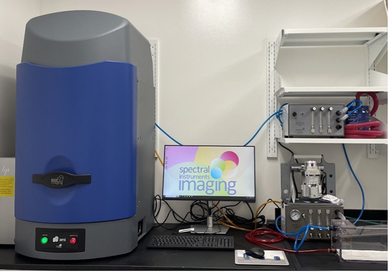
- Lago Spectral Instruments Imaging (room 060E)
- GFP/RFP Panoramic Imaging System (room 060E)
Regular/Transgenic Mice Facility
- Surgery station equipped with isoflurane vaporizer (room 040D1)
- Necropsy station equipped with CO2 chamber (room 040D1)
- Table-top trinocular surgery microscope Meiji EMZ8TR
SCHEDULING
The MCCSAIF has teamed up with MCC Core Information Systems to provide a computer-based method of reserving procedure rooms and equipment. This scheduling system allows researchers to check room availability and/or schedule procedure rooms via the internet.
Scheduling utilizes the University of Kentucky email through Outlook for reservations. Scheduling can be done via the Office 365 online portal or Outlook desktop client.
Reservation rules – maximum 3 hours between 9 a.m.–5 p.m. weekdays.
See below for the available resources.
To schedule Lago SII training please contact:
|
Leif Magnuson |
Stacy Hinchey |

For experimental design questions please contact:
Piotr Rychahou, M.D.
Associate Professor
Markey Cancer Center and Department of Surgery
University of Kentucky
E-Mail: piotr.rychahou@uky.edu
Immunodeficient mice facility
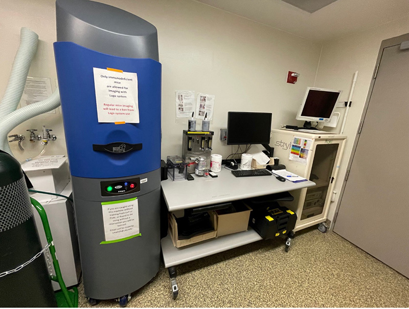 To start using Lago SII imaging system please schedule initial training with Leif Magnuson. Optical imaging is a proven low-cost, high-throughput way to observe tumor growth and treatment effect in living animals. Non-invasive, bioluminescent imaging of tumor growth and metastasis allows longitudinal evaluation of tumor development before, during and after treatment, offering an excellent preclinical strategy to assess tumor response and recurrence.
The Lago SII is connected to the XGI-8 Anesthesia System that allows continuous administration of isoflurane gas for anesthetizing animals during imaging with build in active scavenging system.
Lago SII features:
- Bioluminescent imaging of metastatic tumors (quantification in vivo and ex vivo)
- Detection of fluorescent reagents administered to mice or fluorescent GFP/RFP tumors (signal quantification ex vivo)
- 5 mice imaging across BLI, FLI
- 10 mice imaging across BLI, FLI with available nose cone adapter
- Aura Image Analysis Software is available for free upon request from Spectral Instruments Imaging (SII)
- Analysis of fluorescent reagents distribution in vivo and ex vivo. Recommended dye spectrum for in vivo and ex vivo analysis of fluorescent reagents is Alexa647 or NIR
Note: Markey Cancer Center is supporting use of optical imaging experiment and there is no charge to use the Lago SII. Please not that isoflurane is not provided.
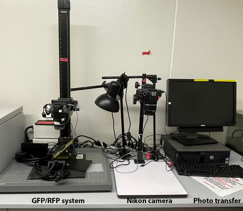 GFP/RFP Panoramic Imaging System
The GFP/RFP Panoramic Imaging System is used for visualizing fluorescent proteins expressed in cancer cells. A major benefit of this imaging system is real-time ex vivo examination of fluorescent signal to determine distribution of metastatic tumors within body cavities (eg, peritoneal cavity lymph node metastasis) and high-resolution photography to demonstrate metastatic burden in organs or body cavities. The detection and demonstration of metastatic tumors is enhanced in metastatic tumors established with cells expressing eGFP or tdTomato RFP enriched with two-three flow-cytometry selection cycles.
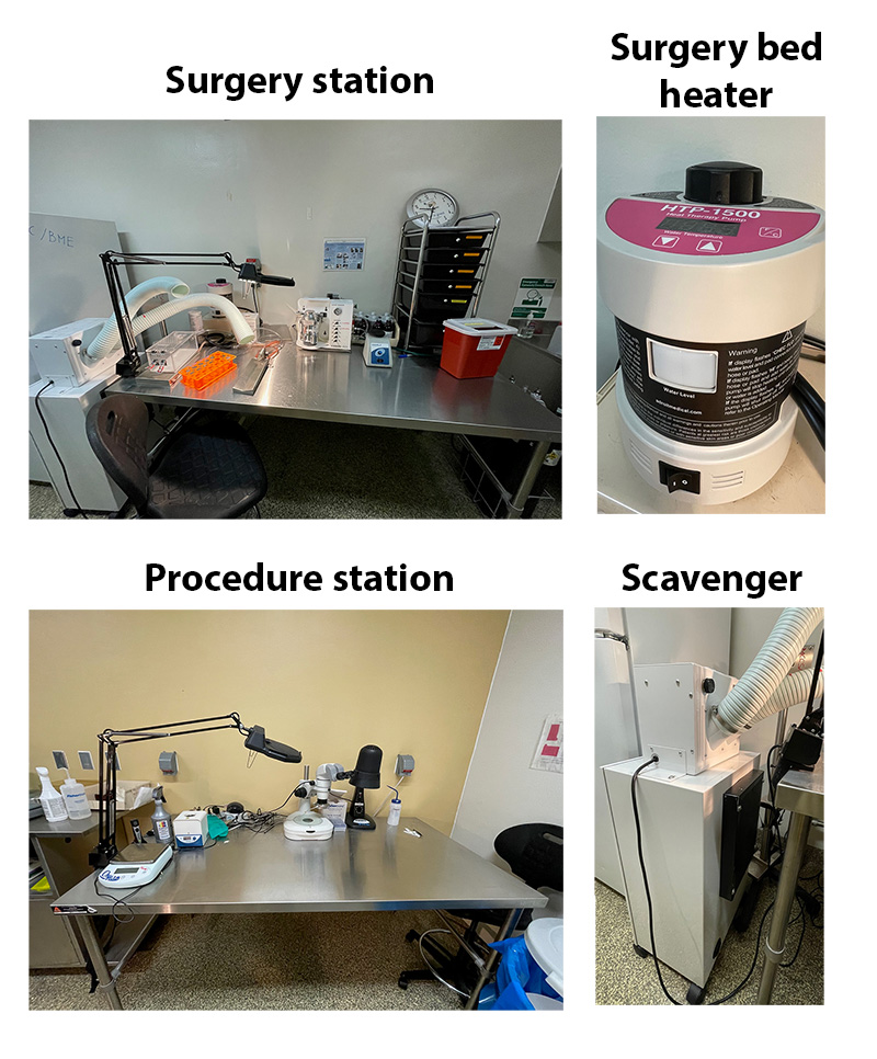 Each surgery station is set up with a dedicated isoflurane vaporizer, heated surgical beds, induction chambers, microflex breathers and a scavenger system. Please follow following procedures:
- Surgery stations are equipped with two scavenging systems – passive, part of an isoflurane vaporizer and active scavenging system.
- Check passive isoflurane vaporizer scavenger system filter weight. Each filter in use is labeled with initial filter weight. If weight exceeds initial filter weight by +50 g discard the filter and replace with a new filter. Measure a new filter weight and write initial weight on a filter.
- Turn on water pump to heat surgical beds. It takes approximately 30 min to heat surgical bed to 42°.
- Turn on active scavenger system and position scavenger system arms over induction chamber and microflex breather.
- Check isoflurane levels and refill if needed. Make sure that oxygen is off and there is no pressure in a vaporizer before refilling isoflurane.
- Turn on oxygen and adjust isoflurane flow.
- Discard syringes used for injection into provided container. Do NOT recap needles.
- Turn off vaporizer, keep the oxygen flow to remove excess of isoflurane though passive scavenging system.
- Turn off oxygen and scavenging system.
- Clean induction chamber and surgical station after each use with provided.
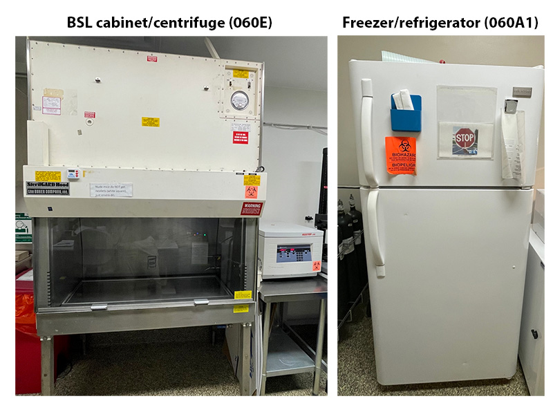 Additional equipment in immunodeficient facility: Nikon camera and stage with light to take gross photographs of organs (060E), centrifuge (060E), BSL cabinet (060E), freezer/refrigerator to store reagents/feed (060A1; must be stored in a labeled container), CO2 chamber (060E)
Regular mice facility
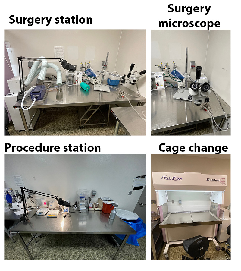 Table-top trinocular surgery microscope Meiji EMZ8TR with zoom stereo on S4200 Boom Stand (zoom range: 7x to 45x), surgery station, procedure station, cage change station, CO2 chamber
partnering facilities
 Partnering Core: Ami HT (Regular mice; Light Microscopy Core, HKRB Room 084B)
https://www.research.uky.edu/light-microscopy-core
The Ami HT small animal imager from Spectral Instruments Imaging is capable of detecting both bioluminescence and fluorescence in vivo and in vitro. The system is available for imaging of immunocompetent rodents. It features 10 excitation wavelengths and allows imaging of up to 5 mice simultaneously. The integrated anesthesia system and mouse friendly platform enable easy and reliable imaging and analysis. The imaging acquisition/analysis software, Aura Imaging Software, provides advanced quantification and analysis of all data, and it is a free software available online for both PC and Mac users.
Please feel free to contact XuFu@uky.edu for any inquires.
MRISC (Magnetic Resonance Imaging and Spectroscopy Center)
https://www.research.uky.edu/magnetic-resonance-imaging-and-spectroscopy-center
In vivo MRI and MicroCT are ideal for serial imaging animals to follow tumor progression. Furthermore, injectable agents can be used to enhance tumor contrast or interrogate metabolic activity. Please contact both David Powell, PhD at david.k.powell@uky.edu and Eric Forman at esform2@uky.edu if you would like to discuss using the MRISC.
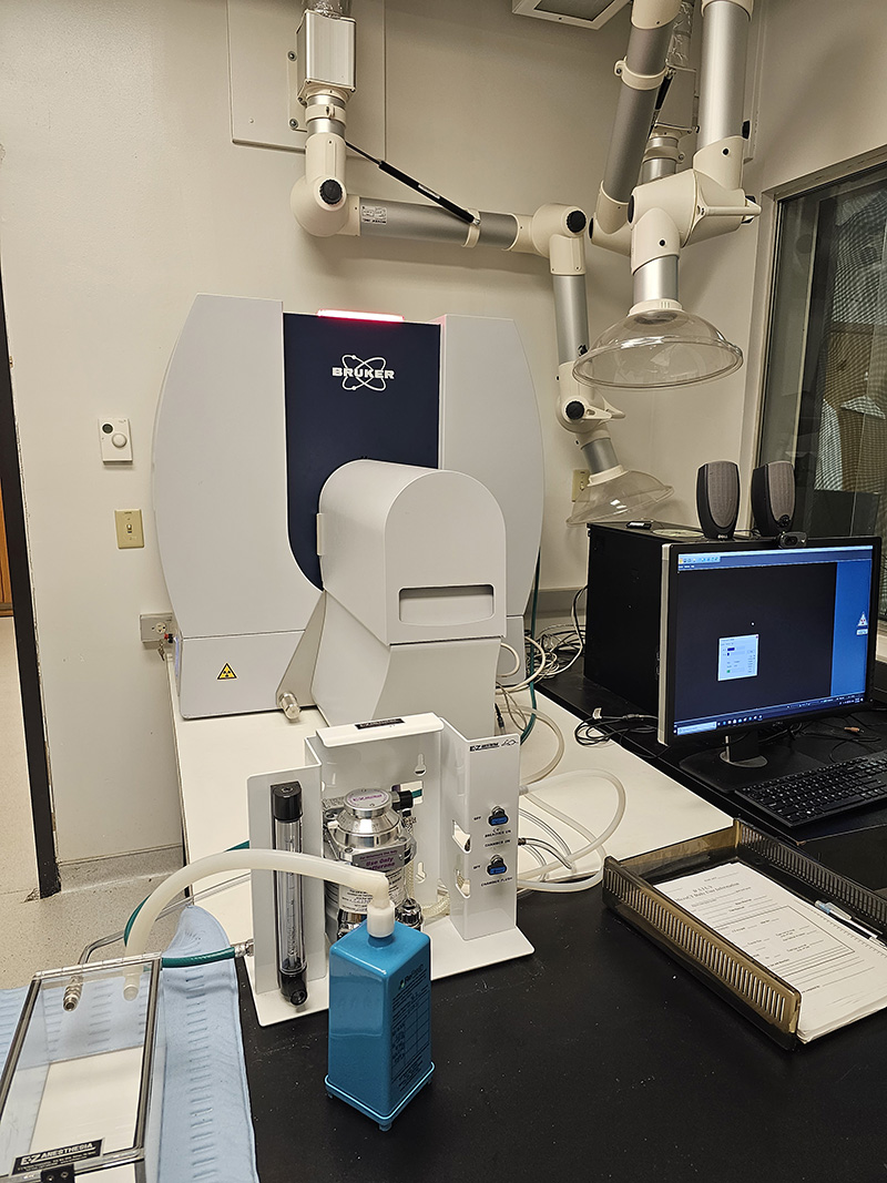 A Skyscan MicroCT 1276 is available at the MRISC basement of the Whitney-Hendrickson building. Usage training and a calendar reservation system are available.
The Bruker SkyScan 1276 is a fast, desktop in vivo micro-CT scanner for small laboratory animals (eg mice and rats) and biological samples. With micro-CT you can non-invasively scan any cross section through the body. Scan bone and tissue samples with high spatial resolution down to 2.8 µm pixel size. Generate realistic 3D images with a 16 Mp CMOS camera, and calculate internal morphology, including specific bone structural parameters.
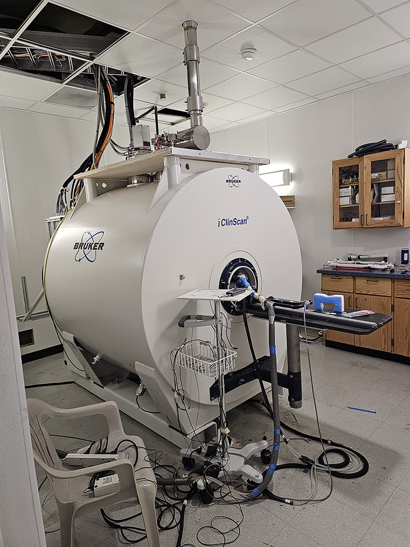 7T BioSpec Scanner. The MRISC is equipped with a new 7T Bruker BioSpec, small animal, MRI scanner. This scanner is ideally suited for translational research with the capability to employ perfusion, diffusion tensor, anatomical, contrast, multinuclear spectroscopy, and functional techniques. We have experience imaging the heart, brain, spine, fat, lungs, vascular system, and cancer.
The highest resolution achievable with the Bruker 1276 is 3 µm. In vivo imaging of mice is limited to around 8 µm. Real-time motion detection and respiratory and cardiac gating are available. Note that 3 µM resolution would require both a significant amount of time to fulfill the S/N requirements and would have to be performed on ex-vivo sample.
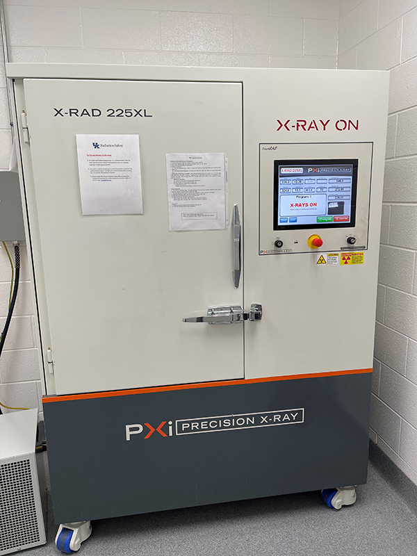 X-Rad225XL (Precision X-Ray Irradiation; PXi) is a system designed for small animal irradiation and large cell cultures. X-Rad225XL is located in the basement of Healthy Kentucky Research Building. The system is under an optional DIY policy; the researchers can irradiate cells and mice by themself. Please read the instructions below on how to proceed.
Please note that the charge to use irradiation is $30/30min/day for cell culture XRT exposures (cell culture, worm, etc.) and $100/1h/day for animal exposure.
Access and training
- Request your access to HKRB 037. Send your request to DLARBusOffice@uky.edu; explain that you need to access HKRB 037 to use X-Rad225XL irradiator. HKRB 037 is located in an animal facility, therefore you will need to perform retina scan with DLAR to gain access.
- Once you have access to the room, contact Dr. Tadahide Izumi t.izumi@uky.edu for a PXi X-Rad225XL specific training. The training normally takes 20 min to complete; during training we will generate PI-specific login account.
- You will also need to complete radiation safety training (contact Gina Carlton (gina.carlton@uky.edu). Complete training with Dr. Izumi before requesting and completing radiation safety training.
- For animal irradiation researchers can reference core protocol 2019-3174 for HKRB 037 location. You may also need to add or update your current protocol by adding HKRB 037 location.
Booking procedure
- Plan radiation procedure between 10am - 5pm.
- Let Dr. Izumi (t.izumi@uky.edu) know in advance what day you want to use the machine.
- Log-in into X-Rad225XL using your account information and proceed with irradiation.