Advanced Cardiovascular Imaging
Cardiovascular imaging is specific imaging studies that allow the physicians in the UK Gill Heart & Vascular Institute to see your heart working inside your body. These images can show the structure, size and shape of your heart in great detail, which can both help with diagnosis of a cardiovascular condition or preparation before a surgery or procedure.
Types of Cardiovascular Imaging
Our advanced cardiac imaging includes cardiac CT scanning, cardiac MRI, cardiac PET and echocardiography. These procedures are noninvasive and essentially painless, requiring only an IV insertion in some instances. All of the imaging can be performed in a comfortable outpatient setting.
- Cardiac CT uses contrast injections to conduct a coronary angiography and to assess your coronary artery calcium scoring.
- Cardiac MRI is the only nonsurgical way to see scarring in your heart after a heart attack and is the most accurate evaluation of heart muscle function. It is often used for preoperative and postoperative evaluation of congenital heart disease.
- Cardiac PET is a nuclear based technique that allows the detection and quantification of myocardial ischemia and blood flow. It can also be used to detect inflammation in conditions such as sarcoidosis and to detect infection in patients with cardiac devices or prosthesis.
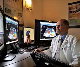
- Echocardiography, or echo, uses high-frequency sound waves to visualize the heart muscle, heart valves (doorways between the heart’s chambers) and blood flow.
Before Cardiovascular Imaging
Depending on the specific procedures you need, you may be asked to limit food and caffeine consumption prior to imaging. If you have kidney disease, you may need blood work to insure you can safely receive the contrast dye. If you are breastfeeding, you should pump and store enough breast milk for two days so your baby will not accidentally consume any contrast. Before an MRI, you will also need to remove all metal from your body, including any piercings.
During Cardiovascular Imaging
You will lie on a special table in the imaging suite, and electrodes will be placed on your chest. If you are having a cardiac CT or MRI, you will be given a contrast injection. It may taste metallic and make you feel flushed. If you are getting an echo, your chest will be rubbed with a gel to improve how the sound waves move through you. You may have to hold your breath at certain points, and you may be given a beta blocker to slow the beating of your heart. An MRI will be very loud.
After Cardiovascular Imaging
Once the imaging has ended, you can get dressed and leave the hospital or go meet with your doctor, depending on what has been scheduled for you. If you were given medication, you may be asked to stay until the side effects have worn off. You should drink plenty of water during the following 24 hours to help expel the contrast from your body, and your urine may be an unusual color.
CONNECT WITH US ON
SOCIAL MEDIA